Categories
Digibaby

How Can Ultrasounds Help to Reveal your Baby’s Gender?

How Can Ultrasounds Help to Reveal your Baby’s Gender?


There was a time when parents waited till birth to know their baby’s gender but this is not the case anymore. Advancement in technology has made it possible for would-be parents to know their baby’s gender before birth. Excitement and liberty to make choices drives parents to go for gender determination. Knowing the gender in advance helps parents better plan things for their baby. Today, the majority of parents want to know their baby’s gender. They have a common motive, but the reason to know their baby’s gender may differ from parent to parent.


A research study performed in 2012 throws more light on these reasons. Four hundred pregnant women undergoing amniocentesis participated in this study. Its findings were published in the journal, Obstetrics and Gynecology.  They were given a questionnaire that asked them why they wanted to know fetal gender after amniocentesis or ultrasound investigation. They gave three reasons for this. 77.8% of the women wanted to know “out of curiosity.” 68% “just wanted to know,” while 66.8% wanted “because it was possible.”

Knowing their baby’s gender in advance helps parents make plans accordingly. They can buy gender-specific clothes and toys. Furthermore, they can choose gender-specific nursery decor. If having other children, they can know which things to keep and which ones to deliver to the charity shop. Additionally, gender knowledge helps parents plan a gender reveal party to enjoy with their near and dear ones.


How can parents determine their baby’s gender?

Ultrasounds

A prenatal ultrasound enables medical experts to identify the gender of a baby growing in the mother’s womb. The ultrasound uses audible sound waves to produce images of a fetus’s shape and position in the uterus. These images display on the monitor. Called fetal anatomy survey, this ultrasound is primarily done to find fetal anomalies. Moreover, it is used for finding the gender of the baby.


Procedure:

You must lie on the examination table in a reclined position for the prenatal ultrasound. Then the ultra-sonographer will apply a gel to your abdomen. Afterward, ultra-sonographer will glide a plastic transducer over the abdomen, emitting high-frequency sound waves. These waves will bounce back after touching your baby’s body and produce an image of the body. The image will display the baby’s soft tissues, organs and other anatomical structures, including reproductive parts. The image will get displayed on the monitor for your viewing.


How do doctors identify your baby’s gender?


The doctors look for certain signs (characteristic features) in the image of the ultrasound to determine the gender of your baby. The signs in boys differ from the signs in girls.


What are these signs?


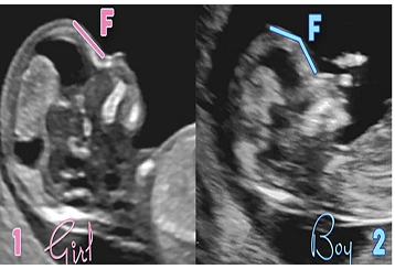
Girl Ultrasound signs:


Hamburger sign: This sign indicates the appearance of the labia and clitoris in an ultrasound. Looking closely at the image, you will see that the labia lips look similar to a hamburger bun. Furthermore, the clitoris will resemble a hamburger patty.


Sagittal sign: Both genders have a sagittal sign. It is obtained by looking at a profile view of the fetus (known as the midline sagittal plane). There is a nub (a small lump or protuberance) at the spine’s end, called a caudal notch. The fetus is a girl if the nub points downward at a 10-degree angle.


Boy Ultrasound Signs:


Male fetuses are not always easier to identify than female fetuses. Identifying male fetuses is difficult before week 14 as the external genitalia has not appeared externally until about 13 weeks. In weeks 18 to 20, a baby boy’s gender can be determined based on the following signs:


Sagittal sign: The fetus is a boy when the caudal notch is pointing upward at more than a 30-degree angle. It can become harder to make a definitive determination if it is somewhere in between.


The flow of urine: The flow of urine can be spotted in a fetus. If the urine is seen moving upward, the baby is likely a boy.


Male genitalia: The presence of male genitalia, including the penis, scrotum and testicles, is a clear sign of male sex. This is often viewed by weeks 18 to 20.


Is gender reveal ultrasound accurate?

Ultrasound - Gener Reveal

The majority of prenatal ultrasounds performed for gender determination are accurate. But errors may occur in identifying gender. The most common mistake is male fetuses are assigned as female. Various factors can impact the accuracy rate of these ultrasounds.


Gestational Age of the baby

Over 99% of ultrasounds performed between weeks 18 and 20 determine the gender correctly. But their accuracy rate can decrease when ultrasounds are performed before week 14.


Position of the baby

If the ultra-sonographer faces difficulty seeing between your baby’s legs, this will affect the ability to determine the baby’s gender. Some fetuses tend to keep their legs together, which impairs gender determination. If the genitalia of the fetus is not visualized, the ultra-sonographer may not be able to examine it perfectly. As a result, he may make mistakes in guessing baby’s gender.


Mother’s body size

If you are overweight or obese, your larger body size can prevent a clear image of your baby. However, an experienced sonographer should be able to obtain a clear picture of women of all sizes.


Multiple babies

If you are pregnant with more than one baby (twins, triplets or more), your babies could hide their siblings. Due to this reason, the gender determination of one or more babies will become more difficult.


The expertise of an ultrasound technician

How experienced and trained ultrasonographers are in their work will impact the accuracy of the gender determination.


Are prenatal ultrasounds safe?   


When conducted by trained technicians for medical purposes, ultrasounds are considered safe. Moreover, they do not use x-rays or radiation. They only use audible and high-frequency sound waves. All ultrasounds, including prenatal ones, are non-invasive and cause no harm to the patients.


Other ways of gender determination


Apart from ultrasounds, the gender of the baby can be confirmed with amniocentesis or chorionic villus sampling (CVS). In the amniocentesis procedure, fluid is extracted from the sac surrounding your baby using a needle and syringe. In CVS, a needle is used to take cells from the placenta.


Both of these procedures carry risks. But they are extremely accurate in making the gender determination. Furthermore, they can return a result as early as week 11 for CVS and week 15 for amniocentesis.


We hope you are well aware of how a prenatal ultrasound can help identify your baby’s gender. So, if you have decided to go for this ultrasound and are looking for a reputed gender reveal ultrasound center then you can opt for DigiBaby. We have specialist medical professionals and ultrasound technicians. Moreover, we have state-of-the-art ultrasound imaging equipment. You will certainly get a safe ultrasound experience and an enjoyable baby viewing experience here. So, get in touch with our experts to seek more information about the ultrasound. Their advice will help you better prepare for your ultrasound and enjoy it fully.

Categories
Digibaby

Pregnancy Ultrasounds for Assessing Your Baby’s Growth Health

Pregnancy Ultrasounds: A Useful Tool for Assessing Your Baby’s Growth and Health

Pregnancy is both a joyous and worrisome time. The discovery of pregnancy brings intense joy to parents, other family members, and friends. But at the same time, it can give rise to worries regarding the health of mother and baby. Pregnancy ultrasounds are the best way to assess the baby’s growth and health and to examine the mother’s reproductive organs. For a safe and worry-free pregnancy ultrasound experience, you must visit a renowned prenatal ultrasonography diagnostic centre in your area. Here a trained and highly qualified sonographic technologist will perform the ultrasound.

How do ultrasounds work

Ultrasound scanning is an imaging technique that uses high-frequency sound waves to produce images of the body’s internal organs. It is also called sonography.   

A transducer or probe emitting ultrasound waves is used to perform an ultrasound. The probe is placed on the skin after coating it with a thin, conductive gel layer. The gel’s coating allows the waves to pass smoothly through the skin. The emitted ultrasound waves are projected on certain body parts which reflect them. Computer software interprets the information from reflected ultrasound waves and turns them into visible images. The info gets formed as per the strength of reflected waves and the time they take to return.  

Ultrasound imaging holds these advantages over other imaging techniques: –

  1. Ultrasounds provide real-time visualization of the fetus or organs. 
  2. They are non-invasive. 
  3. They do away with the need to use ionization radiation known to cause toxic effects on the embryo.
  4. Ultrasounds provide an interactive experience, and this is because they enable the ultra-sonographer to capture different viewing images by moving the probe.   

To assess your baby’s health and growth following ultrasound options are available to you-

2D Ultrasound  

A 2D ultrasound is a traditional ultrasound scanning that is two-dimensional. This scanning sends and receives ultrasound waves in a single plane. The waves that get reflected then provide a flat black and white image of the fetus through that plane. It is a basic image that outlines the organs inside the body. The image obtained is not true to life, but still, it is clear for doctors to detect and diagnose certain medical issues. Doctors use this ultrasound to measure the baby’s head, stomach and leg size. Moreover, they use ultrasound to monitor the heart rate and movement of the baby. In short, a 2D ultrasound is useful for a detailed evaluation of fetal anatomy and morphology.

3D Ultrasound]

3D ultrasounds provide three-dimensional static images of the inside of the body. They are obtained by piecing together multiple two-dimensional images taken at various angles. These ultrasounds visualize fetal structures and the internal anatomy as static 3D images.  

A 4D ultrasound is a 3D ultrasound but in a live motion. It allows doctors to see what is happening inside your body in real-time. For example, it can show how blood flows through the aorta and surrounding blood vessels. You can have a 4D ultrasound during the middle or end of your pregnancy to check for any abnormalities. Also, the ultrasound can show your baby’s activities like yawning, kicking, etc. They will help to provide you with a positive bonding experience.   

Side Effects of Ultrasound 

When used for diagnostic purposes, ultrasound imaging can cause cavitation or small gas pockets in the tissues. Also, ultrasounds can produce slight heating of the tissue. But medical professionals have not witnessed any significant health consequences from their use.

To have a safe and memorable pregnancy ultrasound experience, you need to get it done by a highly qualified California sonographic technologist at DigiBaby. It is an established diagnostic center for pregnancy ultrasonography in California with latest imaging equipment and expert medical professionals. But before booking an appointment to undergo any of the above elective ultrasounds, you need to do one thing. You should consult its medical experts regarding the ultrasound. You should ask them about its benefits, advantages, preparation measures, time spent, etc. The experts will provide you a useful advice about these aspects. Also, they will give you thoroughly enjoyable and safe imaging and baby viewing experience whose memories you will cherish forever.

baby health digibaby

Categories
Digibaby

What Is An NT Scan? Why is it important to perform it between the 11th and 14th Week of Pregnancy?

What Is An NT Scan? Why is it important to perform it between the 11th and 14th Week of Pregnancy?

Discovering that you are pregnant gives rise to a myriad of emotions in you. You may feel intense joy at some moment, while at the other you may feel stressed, anxious or fearful of your baby’s health. You have very little control over your emotions, but you can’t take your baby’s health for granted. So you need to follow up with your doctor from time to time to monitor your and your’s unborn child’s health. Your doctor may recommend specific tests to check if you or your child is affected with a particular health condition.

Some of these tests are in the form of screenings, and these help doctors determine the amount of risk you or your child may have for a specific disease. Screenings can help to identify problems like anemia or gestational diabetes in women. Whereas in unborn children, these can help to detect the presence of chromosomal abnormalities.

The results obtained from these screenings can be positive or negative. A positive result does not ascertain that the child has a chromosomal abnormality. Similarly, a negative result does not guarantee that the unborn child does not have a chromosomal abnormality. Additional diagnostic tests like chorionic villus sampling or amniocentesis must be performed to confirm whether the child has a chromosomal abnormality or not.

What is an NT Scan?

NT Scan, also known as the nuchal translucency test, is a screening test. Non-invasive in nature, it is performed to check the extent of risk (high or low) your child carries for a particular chromosomal abnormality.

NT Scan - Digibaby

NT scan is primarily used for assessing the risk for Down Syndrome and other chromosomal abnormalities like Edwards Syndrome and Patau Syndrome. Also, it helps to determine the risk for some structural problems like major congenital heart problems, abdominal wall and skeletal defects.

Babies born with Down syndrome contain an additional copy of chromosome 21, and this syndrome is incurable and causes developmental delays in them. Such babies have distinct physical characteristics like a small stature, eyes with an upward slant and a low muscle tone.

Edwards syndrome and Patau syndrome are rare chromosomal abnormalities and often prove fatal for babies. Unfortunately, most babies born with these abnormalities die within the first year of their life.

Ultrasounds use high-frequency sound waves to create an image from inside the patient’s body. The nuchal translucency test also uses ultrasound to assess the risk for a medical condition.

All pregnant women are offered the NT scan along with a blood test as a part of first-trimester screening options. The first trimester starts on the first day of the last period and lasts till the end of the 13th week. Apart from the first trimester, screenings are also done in the second and third trimesters.

In an NT scan, a doctor or technician measures the clear (translucent) space in the tissue at the baby’s neck. This translucent space is the nuchal translucency. During the first trimester, babies having abnormalities tend to accumulate more fluid at the back of their neck, and as a result, this clear space becomes larger than average.

Apart from nuchal translucency, some doctors also look for the presence of the fetal nasal bone during the NT scan. If no nasal bone is visible in the developing baby, some experts believe this increases the chance of chromosomal abnormalities in the baby.

What is the right time for performing the NT Scan?

The NT scan must be conducted when a woman is between 11 and 14 weeks pregnant, and this is when the base of her baby’s neck is still transparent. The last day for doing this scan is when a woman is 13 weeks and six days pregnant.

How is the nuchal translucency test performed?

An NT scan requires no special preparation. In most cases, the scan gets completed in just 30 minutes. The doctor will recommend you drink water an hour before the procedure as ultrasound pictures are easier to read with a full bladder. The doctor will ask you to lie down on an exam table to perform the scan. After which, the sonographer will move an ultrasound wand over your stomach. Correspondingly, the pictures of the baby will become visible on the monitor.

The sonographer first confirms the baby’s gestational age by measuring him from crown to rump to see if his size fits the babies of his age. Then he positions the transducer (a sensor) over the abdomen. It shows the baby’s nuchal translucency on the monitor, and the sonographer uses calipers to measure its thickness on the screen.

You can get the results of the nuchal translucency test right away or you may have to wait for ten days if the doctor needs to send the data to a processing center.

How to Interpret NT Scan Results?

The doctor will use a computer program to calculate the amount of risk your growing child carries for a chromosomal abnormality. He will enter your age or date of birth in the program, and it will accordingly give the results.

The results will be in the form of a ratio that expresses the chances of your baby having a chromosomal defect. You will find it confusing to interpret the results yourself. So you are advised to talk to your doctor or a genetic counselor to grasp a better understanding of the results.

The doctor may also tell you that your results are normal or abnormal for a specific condition, depending on whether the ratio is below or above a particular cutoff.

A normal screening result (screen negative) does not guarantee that your child does not have a chromosomal abnormality. But it does suggest that the occurrence of a chromosomal problem is unlikely. Similarly, an abnormal screening result (screen positive) does not mean your baby carries a chromosomal problem, and it just indicates that your baby is more likely to have a problem. The majority of screen-positive babies don’t have a problem. Only one-third of babies with an increased translucency measurement are born with chromosomal defects.

What do doctors consider for calculating NT scan results?

Nuchal translucency measurement is not the sole factor that doctors consider for determining the result of the NT scan. They also consider other factors like the mother’s age, the baby’s gestational age, and the blood test results.

Although chromosomal abnormality can occur in the baby of any mother, the risk of its occurrence increases with the increased age of the mother. For example, if your age is 25 years, your baby’s risk of having Down Syndrome is 1 in 1,200, but if your age is 40 years, this risk increases to 1 in 100. Because of this, doctors consider mother’s age as one of the factors in determining NT scan results.

Is the Nuchal Translucency test accurate?

Like other screening tests, the nuchal translucency test is also not perfect, and it cannot detect all cases of Down Syndrome. The NT test provides both false-negative results and false-positive results.

A false-negative result identifies your baby as a low risk for Down Syndrome while he has this chromosomal condition.

A false-positive result suggests that your child is at increased risk for Down Syndrome or other conditions, but he has no problem.

What to do if your nuchal translucency test result indicates a problem?

After talking with your practitioner or genetic counselor, you can better understand whether your child is at an increased risk for a chromosomal condition.

If there is an increased risk, the next step is to get a diagnostic test like amniocentesis or CVS conducted. The diagnostic test is necessary to confirm a positive screening result for your baby and to obtain a definitive diagnosis.

A needle is inserted through the stomach into the amniotic sac to retrieve a fluid sample for the amniocentesis diagnostic test. Amniotic fluid contains cells that provide genetic information about the baby.

In CVS or chorionic villus sampling, a mother’s placental tissue sample is removed and tested for chromosomal abnormalities and genetic problems.

These diagnostic tests are more invasive and carry a small risk of miscarriage.

Suppose you decide against diagnostic testing. Then you can opt for NIPT, quad screen and detailed ultrasound to know more about your baby’s health and development. The NIPT is a blood test available to all pregnant women at the beginning of 10 weeks of pregnancy. The quad screen is a blood test performed in the second trimester, whereas a detailed ultrasound is performed in 18 to 20 weeks.

A detailed ultrasound can help detect soft markers of chromosomal disorders. Some of these markers are a bright bowel, a bright dot in the heart, short limbs and specific problems in the kidneys. This ultrasound can also be used for finding anatomical defects like spina bifida.

Is it mandatory to undergo the NT scan and diagnostic tests?

It is your discretion to go for the nuchal translucency scan or any diagnostic tests or ignore them altogether. You may choose one, two, or more tests out of all the tests offered to you during the pregnancy and avoid the rest.

By now, you may have understood the need to get the NT scan conducted on your growing baby. If you are keen to get this scan done in Livermore, California, DigiBaby is the place to go. We also provide this facility in Woodridge, Illinois and Mesa, Arizona. Our medical center is equipped with state-of-the-art pregnancy screening, diagnostic equipment, and expert medical professionals to provide you with a safe and satisfying medical experience during your pregnancy. Thus, making your journey to motherhood smooth and enjoyable.

Categories
Digibaby

Importance of prenatal care during pregnancy

Importance of prenatal care during pregnancy

Pregnancy brings a lot of changes to a woman’s body. Women who have been healthy before pregnancy might also experience certain issues during the pregnancy phase. Thus, a woman needs to take proper care, nutrition, supplements after conceiving a baby. Women should focus on proper prenatal care to minimize the risk of complications during and after their pregnancy. Prenatal care is also called antenatal care, and it helps in improving both fetal and maternal outcomes. It helps the doctor properly diagnose, treat, and manage the women’s health conditions before getting serious. Women should always start their prenatal care at the early stage of pregnancy (first trimester) for better health. 

The Importance Of Prenatal Care And Where To Find It - MIBluesPerspectives

Knowing prenatal care in details 

Prenatal care is the medical healthcare that a pregnant woman receives. Regularly going for prenatal check-ups helps know any potential concerns or issues at an early stage of pregnancy and reduces birth complications. It would be best if you visit your physician/gynecologist/obstetrician for screening, diagnostic tests, ultrasounds, counseling, education, emotional and social support, and fetal monitoring. With the help of prenatal check-ups, you can monitor your baby’s growth, heart rate, any signs of premature labor, maternal blood pressure, etc. Doctors will generally schedule your first prenatal appointment when you are at least 8 weeks pregnant. Moreover, the doctor also checks the pre-existing conditions for the mother-to-be, such as diabetes or high blood pressure before pregnancy.

Women having no complications during pregnancy can visit for their scheduled prenatal appointments as follows –

  1. Up to week 28 – 1 prenatal visit per month
  2. Weeks 28 to 36 – 1 prenatal care visit in every 2 weeks
  3. Weeks 36 to 40 – 1 prenatal visit every week

What should you expect from your first prenatal visit?

Prenatal care visit includes tests and general information about what a pregnant woman should and shouldn’t eat or do to stay healthy during their crucial time. The first prenatal visit is often the longest one because the doctor needs to review the entire medical history of the mother and the father. During your first, the doctor would do the following –

  1. Takes blood and urine samples
  2. Records your weight
  3. Checks your blood pressure
  4. Performs a pelvic exam
  5. Review your ultrasound

What will you get from your prenatal appointments? 

  1. 6-12 weeks – As mentioned above, the obstetrician or gynecologist takes your detailed information during the traditional first prenatal visit, including a family history of genetic disorders. They also conduct ultrasound, pelvic exam, blood work, blood pressure, weight, and urine screening for proteins and sugar.12-16 weeks – The second visit is generally suggested after a month of your first appointment. During this visit, the doctor checks the blood pressure, urine screening, and weight recording of the mother and recommends any additional tests. 
  2. 18-22 weeks – During the 18th to 22nd week, ultrasounds are done, and the doctor measures the fundal height (FH) and fetal heart tones (FHTs). Fundal height is the distance in centimeters (cm) from the pubic bone to the top of the uterus. Fetal heart tones include listening and measuring the fetal heart rate with the help of fetal doppler. 
  3. 22-26 weeks – This prenatal visit is almost similar to the last visit, but the only difference is that the doctor also screens the gestational diabetes level of the mother-to-be. The doctor may also check for the swelling in your hands and feet. 
  4. 26-30 weeks – Again, the doctor repeats the measuring of fundal height and fetal heart tones along with the screening for diabetes if not already done. 
  5. 28-32 weeks – At this level of pregnancy, you should visit for prenatal tests frequently as you are getting close to your delivery. FH and FHTs are conducted at every visit to check the growth of your baby. 
  6. 32-36 weeks – At the 36th week of pregnancy, the doctor starts the cervical checks to locate the baby’s position in the womb. 
  7. 36-40 weeks – By now, you must frequently be visiting your doctor, and you’ll be asked to visit every week until the 41st week of your pregnancy. These visits are done to check the mother’s and baby’s health, growth, position of the baby with few additional tests. 

Prenatal care and tests | Office on Women's Health

What is the need for getting regular prenatal check-ups during pregnancy?

Prenatal is essential during pregnancy due to following reasons –

  1. With the help of regular prenatal check-ups, doctors can easily spot pregnancy complications (if any) that may lead to poor fetal growth. Doctors can spot if the baby has any weight issues, neurological problems, etc. So, proper care and attention during pregnancy can reduce the risk of complications for the fetus or baby.
  2. After noting all the factors such as the mother’s age, medical history, etc., the doctor can set up proper tests as per the conditions. It helps control and monitors the mother’s pre-existing medical conditions, such as diabetes, anemia, and blood pressure. Thus, this helps to prevent future complications.  
  3. Prenatal care is also helpful in estimating the delivery date. It puts mothers at ease and helps them to decide on the best plan for her baby.
  4. Antenatal or prenatal care during pregnancy gives a lot of support to the mother-to-be. Proper education and counseling are also part of prenatal care treatment and gives the expecting mother the aid she needs. 
  5. Moreover, proper nutritional advice is also given to the mothers during this crucial stage. A properly balanced diet is essential to prevent several health problems and gives the baby complete nourishment for better growth. Mothers are generally given prenatal vitamins that consist right amount of folic acid and other essential vitamins for baby’s growth.  

Get your regular prenatal visit with Digibaby 3D/4D Ultrasound for a healthy pregnancy 

It is not always too late to seek a doctor’s help during pregnancy. Women who do not go for timely prenatal visit delivers baby with low weight and several birth complications. Every woman needs to stay healthy immediately before, during, and after pregnancy. Thus, it is important to stay updated on all the healthcare appointments as instructed by a doctor.  

You can consult them for 2D ultrasounds, gender determination, and 3D/4D HD live ultrasound as well. All the prenatal care services at Digibaby 3D/4D Ultrasound will be given to you at a genuinely affordable price.

To learn more, contact the center at 877-470-7531 or info@thedigibaby.com now.

Categories
Digibaby

Different types of ultrasound scanning during pregnancy

Different types of ultrasound scanning during pregnancy

Seeing your baby for the first time through a monitor screen can be an emotional and special moment for every parent. Besides taking the first photograph of your baby to your home, ultrasound scanning tests are also vital for many other reasons. The ultrasound scan tests are essential for the doctors to get valuable information such as baby’s growth, detect abnormalities, predict the due pregnancy date, show the position of your placenta, indicate the sex of your baby, etc. These ultrasound scan tests are always performed under the guidance of a trained healthcare professional called a sonographer. Ultrasound scans are performed at different stages of pregnancy for getting all the specific information about the baby.

image

But first, let’s understand the purpose of ultrasound tests

Ultrasound scans are used for getting a sneak peek of your baby during pregnancy and examine how the fetus is developing in the womb. Ultrasound uses sound waves for creating images of your baby along with the images of your uterus, placenta, and surrounding blood vessels and organs. The healthcare professional performing an ultrasound exam uses an instrument called a transducer that sends sound waves through your body. The waves produced are then echoed back to the transducer for transforming them into images. The produced images are then seen on the video screening monitor. With the technological advancement nowadays, 3D and 4D colour scans are also available for ultrasound pregnancy tests for better quality images.

  • It helps in conforming to pregnancy and determining how many weeks pregnant you are.
  • Ascertaining  how many fetuses you are carrying.
  • Confirming the growth of your baby and figuring out your pregnancy due date.
  • It helps to check the abnormalities in your cervix and uterus, if any.
  • Monitors your baby’s heartbeat, muscle tone, movement, and development.
  • Diagnosing any complications during pregnancy such as abnormal bleeding, ectopic pregnancy, molar pregnancy, and miscarriage.
  • Determining the position of the baby.

Different types for pregnancy ultrasound scanning tests

Different ultrasound scanning tests depend on how long your pregnancy is and what the doctor has to check. Here are some common kinds of ultrasound tests –

  1. Transabdominal ultrasound – In this kind of mid-pregnancy scan ultrasound, the doctor uses a transducer for scanning and is the most common ultrasound test. For this, you must lay down on your back on the examination table, and then the doctor covers your belly with a thin layer of gel. The gel applied to the belly helps the sound waves to move easily for a better image. Then the transducer is moved all across your belly. The patient should drink several glasses of water before the transabdominal ultrasound to have a full bladder during the test. A full bladder helps move sound waves easily. The transabdominal ultrasound is a total of 20 minutes’ process.
  2. Transvaginal ultrasound – During a transvaginal ultrasound, a wand-shaped transducer is placed inside your  vagina. This ultrasound test is generally used at the early stages of pregnancy for verifying your pregnancy, checking your baby’s heart rate & position, etc. For this test also, you have to lay down on your back with your feet in stirrups. Then the doctor moves the transducer in your vagina for getting the required information. Your bladder for the transvaginal ultrasound should be empty or just partly full.  
image

Here are some other kinds of ultrasound tests that are done for getting more information about your baby –

  1. Doppler ultrasound – The Doppler ultrasound is useful for checking the baby’s blood flow and the blood flow in your uterus and placenta. It is essential for observing the movement of the blood vessels in your baby. The doctor uses the transducer to listen to the baby’s heartbeat and measure the blood flow in the umbilical cord. Doppler scanning is normally used in the last trimester of your pregnancy.
  2. 3-D ultrasound – This kind of scanning ultrasound test can be done transvaginal or transabdominal. It is used for taking more detailed images of your baby. A 3-D ultrasound test takes thousands of pictures at once and helps in getting a clear 3-D image. Doctors might use this ultrasound to check whether the baby’s organs are developing at a normal pace or not.
  3. 4-D ultrasound – The 4D ultrasound scan is just like the 3D ultrasounds. But the only difference is that it also shows the moving video of the baby’s movement instead of still images. It captures the highlights and shadows for creating a clearer image of the baby’s face and movements. This ultrasound scan can only be requested after 26 weeks of pregnancy.

Consulting the experts for pregnancy ultrasound scanning tests – Digibaby 3D/4D Ultrasound

The ultrasound tests give the highlights of your pregnancy, and it is a truly exciting experience for the parents to see their baby in the mother’s womb. However, you must always trust the experienced professionals who follow all the medical procedures to ensure a healthy pregnancy and safe deliver. Consult the highly trained team at Digibaby 3D/4D Ultrasound now. Their experienced sonographers will keep the mother and the baby safe and inform you of the accurate pregnancy data. You can connect with them to look for affordable and high-quality 2D/3D/4D live ultrasounds. Watch your baby grow, smile, yawn and blink with the latest technology by Digibaby 3D/4D Ultrasound.